How Hearing Works

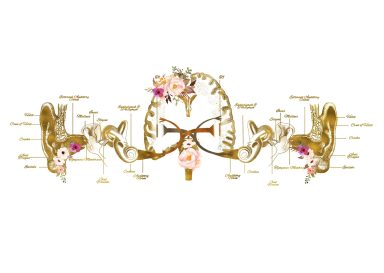
The process of hearing is a complex one, involving several parts of the ear working together to convert sound waves into signals that the brain can interpret. Here's a step-by-step breakdown of how it works:   

1. Outer Ear (Pinna and Ear Canal):

  • The visible part of the ear, called the pinna (or auricle), acts like a funnel, collecting sound waves from the environment.   
  • These sound waves travel through the ear canal (external auditory meatus), a narrow passage leading to the eardrum. The ear canal can also amplify certain frequencies.    

2. Middle Ear (Eardrum and Ossicles):

  • At the end of the ear canal, the sound waves reach the eardrum (tympanic membrane), a thin membrane that vibrates when sound waves hit it.
  • These vibrations are then transmitted to three tiny bones in the middle ear called the ossicles:  
    • Malleus (hammer): Attached to the eardrum.
    • Incus (anvil): Located between the malleus and stapes.
    • Stapes (stirrup): The smallest bone in the human body, connected to the oval window (an opening to the inner ear).
  • The ossicles act as a lever system, amplifying the vibrations from the eardrum and transmitting them to the oval window. This amplification is crucial because the inner ear is filled with fluid, and more force is needed to move fluid than air.   
  • The Eustachian tube connects the middle ear to the back of the throat. It helps to equalize the pressure in the middle ear with the outside air pressure, which is essential for the eardrum to vibrate properly.   

3. Inner Ear (Cochlea):

  • When the stapes vibrates against the oval window, it creates pressure waves in the fluid-filled cochlea, a spiral-shaped structure in the inner ear.   
  • The cochlea contains the basilar membrane, which runs along its length and is covered in tiny hair cells. These hair cells are the sensory receptors for hearing.   
  • Different frequencies of sound waves cause different parts of the basilar membrane to vibrate. High-frequency sounds stimulate hair cells near the base of the cochlea, while low-frequency sounds stimulate hair cells near the apex.   
  • As the basilar membrane vibrates, the stereocilia (tiny hair-like projections) on top of the hair cells bend against an overlying membrane called the tectorial membrane.   
  • This bending of the stereocilia opens ion channels in the hair cells, causing an electrical signal to be generated.   

4. Auditory Nerve and Brain:

  • The electrical signals from the hair cells are transmitted along the auditory nerve (also known as the cochlear nerve or the vestibulocochlear nerve) to the brainstem.   
  • In the brainstem and then the auditory cortex (located in the temporal lobe of the brain), these electrical signals are processed and interpreted as sound, allowing us to perceive pitch, loudness, and other qualities of sound.  


We need your consent to load the translations

We use a third-party service to translate the website content that may collect data about your activity. Please review the details in the privacy policy and accept the service to view the translations.